Home

Microscopy imaging techniques such as confocal and fluorescence microscopy permit visual and spatial investigation of fixed and live biological samples. A light source is used to excite a fluorophore on a target molecule that in turn emits light at a longer wavelength for detection.
Capable of a variety of fluorescence microscopy and image analysis applications, including time lapse experiments. It has YFP, CFP, GFP, Texas Red, Cy5, and A4 filter cubes. It is equipped with the LAS and LAS AF software. Objectives include: Dry (2.5x, 10x, 20x and 40x) and Oil (40x and 60x/100x). The Leica DMI6000 has a colour camera in addition to the fluorescence camera that can be used for bright field, DIC, and phase contrast imaging.
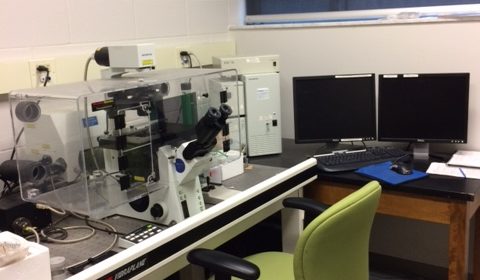
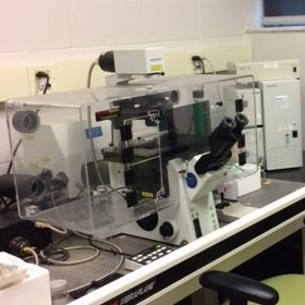
A Biorad 1024 MRC based on a Nikon Eclipse 800 microscope. This unit has the capability of combining double label imaging with DIC transmission optics. It has a krypton/argon laser with excitation at 488 nm, 568 nm and 647 nm. Objectives include 20x, 40x oil and 60x oil. The software includes quantitative analysis for distance staining intensity and double-label co-localization. Triple label sequential analysis is also available. A cryostat (Micron) is available for preparing sections for confocal imaging.
