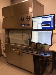
Flow cytometry is a powerful analytical technique fundamental for many cell biology and biomedical applications. Flow cytometry makes use of light from lasers to count and profile individual cell types even, when present in a complex heterogenous cell mixture. This is accomplished by funneling cells through a narrow channel and illuminating the cells as they pass through one at a time. Sensors detect the light that is reflected, diffracted, emitted, and refracted from the cells, while computers are employed to process and analyze this data. Analysis of diagnostic molecular markers are the basis for specific cell type identification and sorting. Among numerous other applications, this technique is able to distinguish disease cells from normal cells within a given tissue sample.
Standard Configuration:
1. Blue ‐ FITC (530/30BP, 505LP), PerCP Cy5.5 (695/40BP, 685LP)
2. Red ‐ APC (660/20BP), AF700 (730/45BP, 695LP), APC‐Cy7/APC‐H7 (780/60BP, 750LP)
3. YG – PE/DsRed (582/12BP), PE‐TR/mCherry (610/20BP, 550LP), PE‐Cy5 (670/30BP, 635LP), PE‐Cy5.5 (710/50BP, 695LP), PE‐Cy7 (780/60BP, 750LP)
4. Violet ‐ BV421/Pac Blue (450/50BP), BV510/AmCyan (528/50BP, 475LP), BV605 (610/20, 595LP), BV650 (670/30BP, 635LP), BV711 (710/50BP, 695LP), BV786 (780/60BP,750LP)
5. UV – Indo‐1 (violet)/AF350/DAPI/Hoechst (450/50BP), Hoechst Red/Side Population (675LP)
Standard Configuration:
1. Blue – FITC (530/30BP 502LP), PerCP‐Cy5.5 (695/40BP 655LP)
2. Red – APC (660/20BP), APC‐Cy7 (780/60BP 735LP), AF 700(760/45BP, 690LP)
3. Violet – BV421/Pac Blue (450/40), BV510/AmCyan (510/50BP, 502LP), BV 605 (610/20BP, 600LP), BV650 (660/20BP, 630LP), BV650 (670/30BP, 635LP), BV711 (710/50BP,
695LP)
4. PE/DsRed (582/15BP), PE‐TR/mCherry, PI (610/20BP 600LP), PE‐Cy5 (670/30BP, 635LP), PE‐Cy5.5 (710/50BP 630LP), PE‐Cy7 (780/60BP 735LP)
5. UV – Indo‐1 (violet)/AF350/DAPI/Hoechst (450/50BP), BUV395 (378/28BP)
Applications
Cell Biology
• Cell Signaling
- Apoptosis pathways
- Cell differentiation
- Cell growth
- Cell proliferation
- Transcription factors
- Cell signaling proteins
• Tumour Characterization
• Fluorescent Protein Detection
• Lineage marker, activation marker, cytokine and chemokine receptors
• Quantification in supernatant, plasma, serum or cell lysates: Cytokines, chemokines, immunoglobulins, or signaling proteins.
• Infection/transduction/transfection confirmation
• Cell tracking
• Small particle analysis
• Functional analysis (ie enzymology, metabolic assays)
• Calcium flux
• Gene expression
• Dye efflux
Cell Viability
• Mitochondrial Analysis
• DNA damage
• Cell toxicity
• Epigenetics and gene regulation
• Autophagy
• Cell death and apoptosis
• Cell cycle
Immunology
• Immunotoxicology
• Human B Cell function
• Chemokine Receptor Expression
Screening
• Antibody quantification
• High-throughput drug screening
Stem Cell
• Characterization and analysis
Microbial Analysis
• Quantification and characterization
Nanoparticle
• Cell uptake
Contact Information:
Facility Manager - Dr. Elizabeth Fidalgo Da Silva Title: Adjunct Professor, Research Associate Institution: University of Windsor Email: fidalgo@uwindsor.ca
Measurement Fees
|